
WETSEMTM Technology
§ High resolution imaging of fully hydrated samples
§ Greatly reduces prep time as well as artifacts
§ Capsule and base fits standard SEMs
§ Safe and simple to use
§ Achieve reliable, reproducible, quantifiable results
Electron microscopy (EM) is today's primary tool for high-resolution imaging the cornerstone for research into living organisms and our material environment.
But because EM requires that samples be placed in a vacuum, it has not been suitable for examining wet samples. This single drawback has been a major impediment to the use of EM for biomedical research, as well as for many other clinical and industrial applications. Until now, the only means of imaging a wet sample with EM was to freeze or dry it, effectively changing it's nature in the process. Such sample preparation is also time-consuming, costly, and often introduces artifacts.
El-Mul (QuantomiX) WETSEM developed with the cooperation of some of the world's leading life sciences and materials science researchers and labs dramatically reduces the preparation needs for high-resolution imaging of wet samples, making imaging easier and faster. It also eliminates many of the artifacts that result when preparing wet samples for EM, opening up opportunities for new research methods as well as new processing methodologies in a variety of fields.
WETSEM also uniquely enables imaging of samples that contain oily and volatile materials.
Also for the first time, WETSEM enables microanalysis using energy dispersive spectrometry (EDS) of wet samples in their native state, a long-awaited capability for many industrial and research labs.
Concept
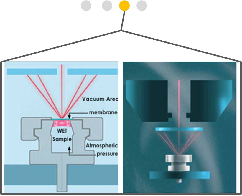
WETSEM Technology is based on a proprietary capsule that is placed on the specimen stage of a standard SEM. At the heart of the capsule is a patented, nanotech-engineered transparent membrane that completely isolates a wet sample from the powerful vacuum used in SEM chambers.
The capsule serves the combined function of specimen holder or cell culture dish, depending on the application. This unique receptacle permits electrons to enter and exit the capsule, while maintaining the sample in its natural state at atmospheric pressure.
Imaging is achieved either with backscattered (BSE) electron detection or X-ray detection, revealing high-resolution structure as well as material composition.
WETSEM is ideally suited for fully-hydrated or liquid-containing samples including foods, oils, cosmetics, ink, biological cells, tissues, and fluid suspensions. In certain settings, WETSEM also enables imaging of dynamic reactions and their products.
Advantages
§ Direct imaging of wet samples (food, cosmetics, inks, cells, tissues)
§ Compositional analysis of wet samples by X-ray microanalysis
§ Wide spectrum of staining and labeling capabilities for cells and tissues
§ Ability to image unstained or unfixed cells and tissues
§ Imaging of both adherent and non-adherent cells
§ High resolution histopathology
§ Intracellular imaging in a scanning EM
§ Imaging the entire cell surface
§ Excellent preservation and imaging of lipid structures
§ Easy-to-automate sample processing and imaging
Capabilities
§ Analyze size distribution, aggregation and homogeneity of particle suspensions.
§ Perform EDS of fully hydrated samples.
§ SEM imaging of oils, greases, volatile substances, emulsions and creams.
§ Enables tissue morphology and analysis.
§ Characterize cellular and subcellular organelles, cell contacts and receptors, cytoskeleton and other detail.
§ Characterize and quantify lipids in fully-wet cells and tissues.
Applications
§ Industrial research: food, oils, dyes, emulsions, pharmaceuticals, suspensions, personal care goods, cosmetics, inks
§ Quality control and quality assurance
§ Life sciences and medicine: cultured and primary cells, histology,nerve cells and myelin imaging, microbiology, plants
§ Tissue implants and prostheses
§ Environmental and toxicological applications
§ Clinical diagnosis: histopathology, cytology, oncology
Recent Publications | |
A. Katz, A. Bentur, and K. Kovlera "A novel system for in-situ observations of early hydration reactions in wet conditions in conventional SEM." Cement and Concrete Research, Volume 37, Issue 1 , January 2007, Pages 32-37. Rossana C. N. Melo, Alon Sabban and Peter F. Weller "Leukocyte lipid bodies: inflammation-related organelles are rapidly detected by wet scanning electron microscopy." Journal of Lipid Research, Vol. 47, 2589-2594, November 2006. Ohad Cohen, et al. "Scanning Electron Microscopy of Thyroid Cells Under Fully Hydrated Conditions - A Novel Technique for a Seasoned Procedure: A Brief Observation." Thyroid Oct 2006. Volume 16, Number 10, 2006: 997-1001. Stephan Thiberge et al. "Scanning electron microscopy of cells and tissues under fully hydrated conditions." PNAS, March 9, 2004. Applied Physical Sciences, Applied Biological Sciences, Volume 101 no. 10. Winston Timp, et al. "Wet electron microscopy with quantum dots." Irit Ruach Nir "A Capsule for Dynamic In-Situ Studies of Hydration Processes by Conventional SEM." Microscopy & Analysis, July 2006. David C. Joy "Scanning electron microscope imaging in liquids - some data on electron interaction in water." Journal of Microscopy, Vol.221, Pt 2 February 2006, pp. 84-88. Federica Boraldi, et al. "Identification of Mineralized Elastic Fibers on Wet Samples by SEM"MICROSCOPY RESEARCH AND TECHNIQUE Volume 67, Issue 6, 2005. Pages 296-299. Guo W, et al. "A new scanning electron microscope method for determination of adipocyte size in human fat tissue: Correlation with weight loss and fat depot burden"poster presentation at the annual meeting of NAASO, the Obesity Society, Vancouver 2005. Abraham Nyska et al. "A new method of wet scanning electron microscopy for the analysis of myelination in EAE mouse model of multiple sclerosis" EXPERIMENTAL AND TOXICOLOGIC PATHOLOGY, November 2005. Irit Ruach-Nir "An Innovative Method for Imaging and Chemical Analysis of Wet Samples in Scanning Electron Microscopes" MICROSCOPY TODAY, July 2005. Vered Behar "Applications of a Novel SEM Technique for the Analysis of Hydrated Samples"Microscopy & Analysis, Issue 108, July 2005. Iris Barshak et al. "Wet SEM: A Novel Method for Rapid Diagnosis of Brain Tumors" Ultrastructural Pathology, 28:255-260, 2004. Stephan Thiberge et al. "An apparatus for imaging liquids, cells and other wet samples in the scanning electron microscope." Review of Scientific Instruments, Volume 75 no. 7, July 2004. Abraham Nyska et al. "Electron Microscopy of Wet Tissues: A Case Study in Renal Pathology."Toxicologic Pathology, Volume 32 no. 3, May 2004. Iris Barshak et al. "A Novel Method for 'Wet' SEM." Ultrastructural Pathology, Volume 28, Number 1 / January-February 04. Opher Gileadi et al. "Squid Sperm to Clam Eggs: Imaging Wet Samples in a Scanning Electron Microscope." Biol. Bull. 205: 177�179. October 2003, Marine Biological Laboratory. |